Cryo-electron tomography

Okay kiddo, have you ever played with a 3D puzzle or looked at your bedroom through a magnifying glass? Cryo-electron tomography is kinda like that, but instead of a puzzle or a magnifying glass, scientists use really powerful microscopes to look at teeny-tiny objects that are too small to see with our eyes.
First, they take a sample of the thing they want to study – it could be a virus, a cell, or even a single molecule. Then, they freeze it REALLY fast – like, -180 degrees Celsius! This freezes everything in place, so nothing moves around while they're trying to look at it.
Next, they chop the frozen sample into thin slices – kinda like slicing bread! But instead of using a knife, they use a fancy machine called a cryo-ultramicrotome to slice the sample really, really thinly.
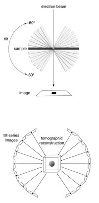
Then comes the fun part – the scientists put the thin slices under a special microscope called a cryo-electron tomography microscope. This microscope takes really detailed pictures of the sample from all different angles, kind of like if you took a bunch of pictures of your toy from every side and put them together to make a 3D picture. Except, instead of just taking pictures from the outside, the microscope looks at the inside of the sample too!
Finally, the scientists use a computer to combine all the pictures into a 3D image – just like putting all the pieces of your puzzle together or looking at your room through the magnifying glass. This lets them see the sample in incredible detail, and they can even see things like individual atoms or molecules!
So, cryo-electron tomography is a really fancy way for scientists to take 3D pictures of super tiny things using special microscopes, computers, and freezing techniques. Pretty cool, huh?
Related topics others have asked about:
